WELCOME TO THE TECMEDLAB TECHNOLOGIES WEB PAGE (still in
progress).
The TECMEDLAB laboratory is fully equipped from the
in-vitro to the in-vivo state-of-the-art technologies for
electromechanical studies in cardiac tissue
Hereafter a brief explanation
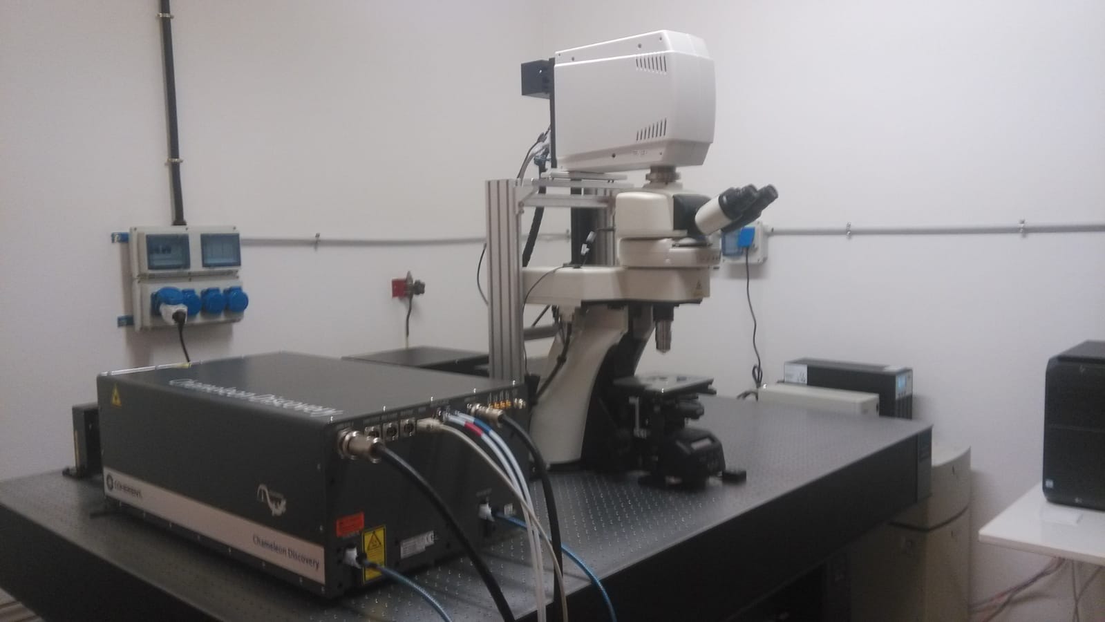
1) THE MULTIPHOTON MICROSCOPY (2020)
The new multi-photon microscopy installed in
2020, is now in place and running in our diffuse
ParmaPhotonics lab!. The system is equipped with
Chameleon Discovery Laser, Nikon Upright Ti Microscope,
fast-resonant scanner (15000 lines x sec.), 4 VIS detectors, 1
spectral detector.
Mux (abbreviation for Multiplexing Epicardial
Mapping) is a system capable to record 16X16 -in parallel-
Electrograms signals at 8-32 KHz temporal Resolution.
Customized together with Crescent Electronics,MUX
is a versatile instrument for in-vivo and ex-vivo (Langerdorff System)
electrograms acquisition and stimulation for epicardial spatiotemporal
mapping of impulse propagation
3) THE Vi.Ki.E. (2016)
ViKiE (Video Kinematic Evaluation) of cardiac
contraction is a novel technology entirely developed in the
Tecmedlab. Is capable to acquire tissue deformation during cardiac
beating and analyzed kinematic parameters and tissue compliance.
What we can do with that: ViKIE is employed in both
preclinical and clinical levels. Preclinical: estimated cardiac
performance in situ or ex-vivo (Langerdorff system) and is totally
combined with MUX for the acquisition of local ElectroMechanical
delay. Due to the high spationtemporal resolution (pixel size 2.5
um, max fps= 1700 ) ViKiE can detect minimal tissue deformation and
analyze the Energy expenditure directly linked to the ATP
consumption. Clinical: ViKiE is employed in the cardiac theater
(thanks to the collaboration with heart surgery in University of
Verona) for measuring in real time kinematics before and after
operation. To now ViKiE has been assessed for Tetrology of Fallot,
CABG, Hypoplasic heart, Heart transplant and pulmonary valve
replacement.

4) The LOKI (2021)
LOKI (Longitudinal OptoKinematic Incubation) is the new-entry
technologies in the TECMEDLAB. Developed in the LabVieW
environment, LOKI consists in an epifluorescence microscope
equipped with three different LED sources, placed in the Incubator
where an high-res videocamera is placed on the side-port.


What we can do with that: LOKI is capable to measure
long.term kinematics (hours, days, months) in beating iPSC-CM, 3D
organoids, neonatal and adult cardiomyocytes. Due to its nature LOKI
can do times laps video recording in both bright-field and
fluorescence for assess cell maturation, metabolism, action
potential propagation, calcium transient, ATP, NOS, ROS etc. LOKI
can also be employed for optogenetic and chemogenetic, thanks with
the possibility to the built-in LEDs and optical fiber for light
stimulation. THe final aim of LOKI is to explore several multiverses
of beating cells from 2D to 3D organoids on a chip.